Overview
 Morton neuromas are focal areas of symptomatic perineural fibrosis around a plantar digital nerve of the foot. The abnormality is non-neoplastic and does not represent a true neuroma. It may more correctly be known as Morton?s metatarsalgia. The condition is thought to be due to chronic entrapment of the nerve by the intermetatarsal ligament. It most often occurs in middle-aged individuals and is many times more common in women than men. Approximately 30% of asymptomatic middle-aged persons have the radiological pathologic findings of a Morton?s neuroma.
Causes
Wearing shoes that are too tight can make the pain of Morton’s neuroma worse. This is because the toe bones are more likely to press on the affected nerve if your shoes are too tight. High-heeled shoes, particularly those over 5cm (2 inches), or shoes with a pointed or tight toe area, can also compress your toes and make the pain worse. This is why women tend to be affected by Morton’s neuroma more than men.
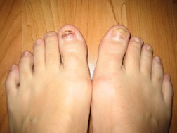
Foot conditions.
Symptoms
The symptoms of a Morton’s neuroma are classic in nature. The patient complains of a burning , tingling, slightly numb feeling (dysesthesias) which radiates out to the toes on either side of the interspace that is involved. For instance, a Morton’s neuroma of the third interspace will result in pain between the third and fourth toes, and a neuroma in the second interspace will cause pain between the second and third toes. The symptoms are usually aggravated by wearing shoes, particularly those with high heels. Symptoms are relieved by walking in flat, wide shoes or going barefoot. Rarely will the patient experience pain when sitting or laying down.
Diagnosis
You might first seek advice from your family doctor about your foot pain. He or she may refer you to a doctor or surgeon who specializes in foot disorders. Before your appointment, you may want to write a list of answers to the following questions. When did your symptoms begin? Did your symptoms begin gradually or suddenly? What type of footwear do you wear for work? Do you participate in sports? If so, what types in particular? What medications and supplements do you take regularly? Your doctor may ask some of the following questions. Is the pain worse in certain pairs of shoes? Does any type of activity ease the pain or worsen it? Are you having pain in any other part of your body?
Non Surgical Treatment
To help relieve the pain associated with Morton’s neuroma and allow the nerve to heal, consider the following self-care tips. Take anti-inflammatory medications. Over-the-counter nonsteroidal anti-inflammatory medications, such as ibuprofen (Advil, Motrin IB, others) and naproxen (Aleve), can reduce swelling and relieve pain. Try ice massage. Regular ice massage may help reduce pain. Freeze a water-filled paper cup or plastic foam cup and roll the ice over the painful site. Change your footwear. Avoid high heels or tight shoes. Choose shoes with a broad toe box and extra depth. Take a break. For a few weeks, reduce activities such as jogging, aerobic exercise or dancing that subject your feet to high impact.
Surgical Treatment
For severe or persistent pain, you may need surgery to remove the neuroma. Once the nerve is gone, you permanently lose feeling in the affected area. One alternative to surgery is to undergo neurolysis injections. These use chemical agents to block pain signals. Another alternative is to take a prescription pain reliever that alleviates nerve pain.
Prevention
Wearing shoes that fit properly and that have plenty of room in the toe area may help prevent Morton’s neuroma.


 Overview
 Symptoms
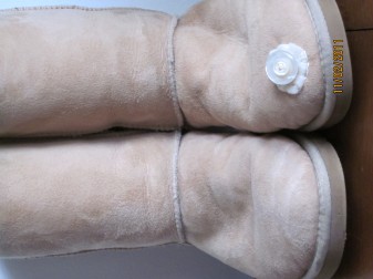 Prevention
 Overview
 Symptoms


 Achilles tendonitis is a condition wherein the Achilles tendon, at or near its insertion to back of the heel, becomes inflamed and causes pain. The Achilles tendon is one of the longest and strongest tendons in the body. It is avascular (not supplied with blood vessels) so it can be slow to heal. The Achilles tendon is formed in the lower third of the leg. Two muscles join to form the Achilles tendon, the Gastrocnemius and the Soleus which are commonly referred to as the calf muscle. The Achilles tendon works as an anti-pronator which means it helps to prevent the foot from rolling inward.
